Similar
American journal of physiology (1898) (14779957994)
Summary
Identifier: americanjourn01ameruoft (find matches)
Title: American journal of physiology
Year: 1898 (1890s)
Authors: American Physiological Society (1887- ) American Physiological Society (1887- ). Abstracts of papers presented at the fall meeting American Physiological Society (1887- ). Proceedings
Subjects: Physiology Physiology
Publisher: (Bethesda, Md., etc.) American Physiological Society (etc.)
Contributing Library: Gerstein - University of Toronto
Digitizing Sponsor: University of Toronto
Text Appearing Before Image:
forced into the trephine hole in the case of the former, thus block-ing off the manometer. Exp. 4. Small dog — bled to death from carotid. Irrigation liquid was bloodof young calves carefully filtered. This blood had been kept over night and hadfrozen. This fact probably accounts for the unusually rapid diminution in flow asthe experiment proceeded, the red corpuscles not passing readily through thecapillaries and clogging them. Inflow cannulas in the subclavians, ligatures beingso placed as to leave an open path only to the vertebrals ; outflow cannulas in thesuperior cerebral veins at their emergence from the skull. 62 /^^. H. Howell. Arterial pressure 60 mm. mercury = outflow not measurable with accuracy. u u igo ■ = of 28.08 c.c. per min. 320 = 47-38 2d series on the same animal. Arterial pressure of 60 mm. mercury = outflow not measurable with accuracy. u ^. 400 = of 42.12 c.c. per min. u u i. ^00 , = 50.02 nininilllllllllHIIUIUHIIIIHIWlllllllllMIUnHIIIUlHIIIIIIlUlllUlMMIMI^^
Text Appearing After Image:
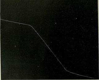
Fig. I. Record of venous outflow from brain under arterial pressures of 60 mm.,380 mm., and 60 mm. Under 60 mm. Hg. the outflow = 18.13 c.c. per min. 380 = 102.66 Return to 60 = 10.24 The time record at the top of the illustration is in seconds. It is evident from the data given that in all the experiments made,the blood-flow through the brain diminished as the experiment pro-ceeded, and this effect was most marked after the blood vessels hadbeen submitted to very high pressures. The probable explanation ofthis fact is that the dead capillary walls permitted a rapid filtrationof liquid, which rendered the brain oedematous. This condition, in- High Arterial Pressures upon the Blooei-flow. 63 deed, was apparent to the eye when the brain was exposed aftersubmission to the high intravascular pressures. This variation fromnormal conditions does not, however, affect the value of the experi-ments so far as the main point under investigation is concerned. The
Tags
Date
Source
Copyright info


