Similar
Comparative embryology of the vertebrates; with 2057 drawings and photos. grouped as 380 illus (1953) (20677416851)
Summary
Title: Comparative embryology of the vertebrates; with 2057 drawings and photos. grouped as 380 illus
Identifier: comparativeembry00nels (find matches)
Year: 1953 (1950s)
Authors: Nelsen, Olin E. (Olin Everett), b. 1898
Subjects: Vertebrates -- Embryology; Comparative embryology
Publisher: New York, Blakiston
Contributing Library: MBLWHOI Library
Digitizing Sponsor: MBLWHOI Library
Text Appearing Before Image:
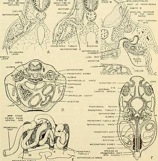
STAGE m Hi, SHORTLY BEFORE HATCHING ELENCEPHALON ROOT OF VAGUS NERVE SOMITE n GANGLION NODOSUM OF THE VAGUS NERVE
Text Appearing After Image:
PRONEPHRIC TUBULE Fig. 346. The developing pronephric kidney in the frog, Rana sylvatica (A-C and E, redrawn from Field, 1891, Bull. Mus. Comp. Zool. at Harvard College, vol. 21. E con- siderably modified). (A) Transverse section through developing second pronephric tubule of frog embryo at a time when the neural tube is completely closed, two gill fundaments are present and the otic vesicle is a shallow depression. (B) Same tubule at about the time of hatching. (C) Section through first pronephric tubule at 8 mm. stage. (D) Transverse section through second pronephric tubule, see line d, fig. 346F, of 18 mm. Rana pipiens tadpole. (E) Entire pronephric kidney of one side of 8 mm. R. sylvatica embryo. (F) Schematic reconstruction of 18 mm. R. pipiens tadpole look- ing down from dorsal area upon the pronephric kidneys and the developing mesonephric kidneys, 779
Tags
Date
Source
Copyright info


