Similar
Gynaecology for students and practitioners (1916) (14781275125)
Summary
Identifier: gynaecologyforst00eden (find matches)
Title: Gynaecology for students and practitioners
Year: 1916 (1910s)
Authors: Eden, Thomas Watts, 1864-
Subjects: Gynecology Gynecology
Publisher: New York : Macmillan
Contributing Library: Francis A. Countway Library of Medicine
Digitizing Sponsor: Open Knowledge Commons
Text Appearing Before Image:
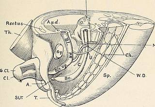
lantoic duct; B., bowel; Ch.,notachord ; Cl.M., cloacal membrane ; C, coelom (future pouch of Douglas) ;G.P., genital papilla ; S.Ur., urorectal septum ; Sin. Ug., sinus urogenitalis ;Sp., spinal cord ; T., tail. (see Fig. 59, M.D.), and produced Miillers prominence on its dorsalwall ; through this they open and their point of opening lies abovethat of the Wolffian ducts. DEVELOPMENT OF GENITOURINARY ORGANS 83 The bowel has been completely separated from the urogenitalsystem by the urorectal septum (.see Figs. 58 and 59). The upper partof the ventral cloacal segment has become the bladder and urachus.The sinus urogenitalis is at this stage an extremely long pouch(see Figs. 58 and 59, Sin.Ug.). Changes in the Cloacal Membrane. Before describing the forma-tion of the external genitals we must consider what has been goingon with regard to the cloacal membrane. This membrane, which reached from the navel to the tail, hasbeen pushed inwards and invested by a thick mass of tissue which W.B.
Text Appearing After Image:
M.D, Fig. 59. Model of the Pelvic Region of a Human Embryo 29 mm. inLENGTH (after Keibel). A, Anus ; A.u.d., art umbilicalis dextra ; Bo., bowel;BL, bladder; C, coelom (pouch of Douglas) ; Ch., notachord ; CI., clitoris ;G.Cl., glans clitoridis ; L., liver; Lig. R., round ligament; M.D., Miillersduct; O., ovary; P.R., primary urethra; Sin. ug., sinus urogenitalis;Sy., symphysis ; Sp., spinal cord ; U., ureter ; Th., thigh ; T., tail; W.B.,Wolffian body ; W.D., Wolffian duct; S.Ur., urorectal septum. forms a ventral papilla (see Figs. 57 and 58, G.P.). This cloacal orgenital papilla partly covers the cloacal membrane. Partly throughthe growth of the genital papilla and partly through two lateralthick swellings—the genital folds—the cloacal membrane is pushedsomewhat into the depths (backwards) (see Fig. 58, Cl.M.). By theunion of the urorectal septum with the cloacal membrane the latteris divided into urogenital membrane anteriorly, and anal membraneposteriorly. The former is the f
Tags
Date
Source
Copyright info


![Secunda pagina figurarum capitalium. [Human brain, 5 pictures] Secunda pagina figurarum capitalium. [Human brain, 5 pictures]](https://cache.getarchive.net/Prod/thumb/cdn6/L3Bob3RvLzE1NDUvMTIvMzEvc2VjdW5kYS1wYWdpbmEtZmlndXJhcnVtLWNhcGl0YWxpdW0taHVtYW4tYnJhaW4tNS1waWN0dXJlcy0yYWNkNjUtNjQwLmpwZw%3D%3D/40/55/jpg)


