Similar
Hand-book of physiology (1892) (14742292056)
Summary
Identifier: handbookofphysio00bake (find matches)
Title: Hand-book of physiology
Year: 1892 (1890s)
Authors: Baker, W. Morrant, (William Morrant), 1839-1896 Harris, Vincent Dormer Kirkes, William Senhouse, 1823-1864. Hand-book of physiology. 13th ed
Subjects: Physiology Human physiology
Publisher: London : John Murray
Contributing Library: Francis A. Countway Library of Medicine
Digitizing Sponsor: Open Knowledge Commons and Harvard Medical School
Text Appearing Before Image:
Fig. 281.—Malpighian capsule and tuft of capillaries, injected through the renal arterywith coloured gelatin, a, glomerular vessels; b, capsule; c, anterior capsule ; d,glomerular artery; e, efferent veins; /, epithelium of tubes. (Cadiat.) CH. X.) THE BLOOD-SUPPLY OF THE KIDNEY. 421 vein. These small veins pass into others which form venousarches corresponding to the arterial arches, but which are moredistinct, situated between the medulla and cortex. Thus, in the kidney, the blood entering by the renal artery,traverses two sets of capillaries before emerging by the renal vein,an arrangement which may becompared to the portal system PS in miniature. The tuft of vessels in thecourse of development is thrust
Text Appearing After Image:
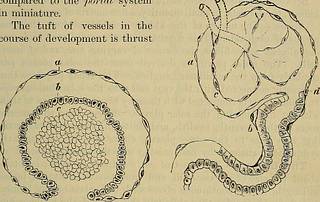
Fig. 282.—Transverse section of a deve-loping Malpighian capsule and tuft(human), x 300. From a foetus atabout the fourth month; a, flattenedcells growing to form the capsule;b, more rounded cells ; continuous withthe above, reflected round c, and finallyenveloping it; c, mass of embryoniccells which will later become developedinto blood-vessels. (W. Pye.) Fig. 283.—Epithelial elements of a Malpi-ghian capsule and tuft, with the com-mencement of a urinary tubule showingthe afferent and efferent vessel; a, layerof flat epithelium forming the capsule ; b, similar, but rather larger epithelialcells, placed in the walls of the tube; c, cells, covering the vessels of the capil-lary tuft; d, commencement of thetubule, somewhat narrower than therest of it. (W. Pye.) into the dilated extremity of the urinary tubule, which finallycompletely invests it. Thus the Malpighian capsule is lined by aparietal layer of squamous cells and a visceral or reflected layerimmediately covering the vascula
Date
Source
Copyright info


