Similar
The American journal of roentgenology, radium therapy and nuclear medicine (1906) (14571151597)
Summary
Identifier: americanjournroen07ameruoft (find matches)
Title: The American journal of roentgenology, radium therapy and nuclear medicine
Year: 1906 (1900s)
Authors: American Radium Society American Roentgen Ray Society
Subjects: Radiotherapy X-rays
Publisher: Springfield, Ill. C.C. Thomas
Contributing Library: Gerstein - University of Toronto
Digitizing Sponsor: University of Toronto
Text Appearing Before Image:
aratively more elevated and ofdarker color than those of the gluteal region.The patches showed a tendency to peripheralextension. Nodules around the joints wereof a rather firm consistency, covered by pig-mented epithelium. Over fleshy areas theyappeared softer, covered with epithelium ofthe same nature, some forming largeplaques as mentioned. Some of the tumorsoccurring over the joints were very hardand seemed attached to the bones. The lesions present in this case were,briefly: I. Nodules of evelids. Case of Xanthoma Showing Multiple Bone Lesions 483 2. Nodules around joints, hands, elbows,knees, and feet. 3. Flat tumors over soft tissues.The palms and soles were free. The interesting features were the differ-ent types of lesion-, soft and moderately high fat diet showed tremendous increasein cholesterol. Fat lower than before. Multiple xanthomata of hand; excision. Several tumors of left hand excised; oneover metacarpophalangeal joint of indexfineer found to involve extensor tendon
Text Appearing After Image:
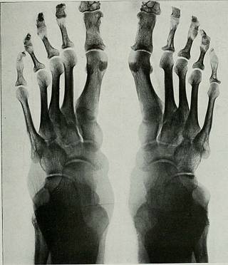
Fig. 2. Symmetrical Lesions in ist and 2Ni> Phalanges ist Toes, ist Phalanges 2nd and 5TH Toes and sth Metatarsals. hard, covered by pigmented epithelium, veryhard ones covered by normal epithelium sug-gesting exostoses, marked symmetry, occur-rences in locations where there was liabilityto friction or pressure, absence of joint in-volvement. Three months later, report of blood after which was trimmed down to normal dimen-sion. Joint itself not involved. Three weekslater excision of tumor of left hand andfingers. Pathological Report.—Elbow: Hard yel-low nodular tumor covered with skin andcomposed on section of spherical firm yellov^r 484 A-Ray nodules 19 x 18 mm., banded together byconnective tissue. Microscopic examinationshowed tumor composed of fibroblasts form-ing a mass of network and containing manylarge vacuolated cells resembling endothelialleukocytes. There were notable light spacesbetween the fibrils which probably containedcholesterin crystals. About these spaces weres
Tags
Date
Source
Copyright info


